
The placenta (from Latin «tortilla», meaning placenta) is a children’s place-fetal organ of all the placental mammals, some marsupials, hammerhead fish, and other viviparous fish. Also, the viviparous Onychophora and other groups of animals transfer the material between the circulation systems of the fetus and mother through the placenta. In botanic placenta is a carpel section to which the ovule attaches.
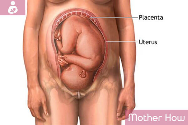
In mammals the placenta forms from embryonic fetal membranes (villous, CVS, and the urinary bag – allantois) which are attached to the walls of the uterus and form the outgrowths (villi), jutting into the mucosa and forming a close link between the embryo and the mothers’ body. This connection serves for the nutrition and respiration of the embryo. The umbilical cord connects the fetus and placenta. The placenta with fetal membranes (so-called afterbirth) comes out of the reproductive tract of the woman in 5-60 minutes (depending on the tactics of the childbirth) after the birth of the child.
Human Placenta
Human placenta (placenta discoidalis) – placenta of the hemochorial type, maternal blood is circulating around the thin villi, which contain fetal capillaries. Since the 30s, the medicine containing extract of the placenta and placenta suspension (developed by professor V.P. Filatov.) is manufactured. Those kinds of medicine are popular enough in pharmacology. The stem cells that are kept in blood banks can be extracted from the umbilical blood and placenta. Placental extracts are also antibacterial and antiviral. The medicine also provides the body with all necessary substrates (vitamins, amino acids) which allow to stimulate the body without depletion of its energetic, plastic, and other resources.
The presence of amino acids, enzymes, and unique protein regulators allows drugs containing the placenta to activate cells of a grown body. That leads to their reproduction, renewing of the cellular composition, and finally to rejuvenating. There is a tradition in some countries to take the placenta home and dig it under the tree. The placenta was considered sterile till it was proved according to the results of research of the project «Microbiome of the human» that its microorganisms are similar to those in the mouth cavity of females.
Animal Placenta
There are several types of the animal placenta. Marsupials have an incomplete placenta which causes such a short period of pregnancy (8-40 days). Cloven-hoofed animals have placenta diffusa of the epitheliochorial type. Placenta Zonaria of endoteliochorial type is observed in predators. Placenta discoid (hemochorial type) in rodents and human and placenta cotyledonaria or multiplex placenta- in ruminants. Most female mammals, including herbivores (cows and other ruminants), eat their afterbirth right after licking the newborn. It is done not only to eliminate the blood smell that may attract predators but also provide them with vitamins and nutrients they need after giving birth.
The Formation and Structure of the Placenta
The placenta is formed of the endometrium and cytotrophoblast in the mucous membrane of the posterior wall of the uterus.
The layers of the placenta (from fetus to the uterus-histologically):
- Decidua-transformed endometrium (with decidual cells rich in glycogen)
- Rohr fibrinoid (Lanthanses layer)
- Trophoblast covering the gaps and growing into the walls of spiral arteries, preventing their reduction
- Lacunas filled with blood
- Syncytiotrophoblast (multicore symplast covering cytotrophoblast)
- Cytotrophoblast ( separate cells forming syncytia and secreting biologically active substances)
- The stroma (connective tissue containing blood vessels, Kashchenko-Gofbauera cells – macrophages)
- Amnion (placental synthesize amniotic fluids, non placental-absorbing)
The Decidua membrane and the socket with maternal blood are placed between the fetal and maternal parts of the placenta-basal. This part of the placenta is divided into 15-20 cup-shaped spaces (cotyledons) by the decidua septum. Each of them comprises the main branch, consisting of the umbilical blood vessels of the fetus.
Those branch out further in a number of chorionic villi that make up the surface of the cotyledon. Due to the presence of a placental barrier the blood flow of the mother and fetus is not connected. Exchange of materials is performed with the help of diffusion, osmosis, or active transporting. From the third week of pregnancy, when the heart of the child starts beating the fetus is supplied with oxygen and nutrients through the placenta.
The forming does not have a clear structure till the 12 weeks of pregnancy. Before the 6th week of pregnancy, it is placed around the ovum and is called the chorion. Placentation takes place on 3rd – 6th week.
Placenta Functions
The placenta forms the barrier, which is represented by the layer of vascular endothelial cells, their basal membrane, a layer of loose connective precapillary tissue, the basal membrane of trophoblast, layers of cytotrophoblast, and syncytiotrophoblast. The vessels of the fetus branching in the placenta to the smallest capillaries form (accompanied with supporting tissues) chorionic villi, which are immersed into the gaps filled with maternal blood. Thus placenta performs the following functions :

Gas Exchange
The oxygen from maternal blood enters the fetal blood according to simple laws of diffusion. Carbon dioxide is transported back.
Trophic and Excretory
The fetus receives water, electrolytes, nutrients and minerals, vitamins through the placenta. Also, the placenta takes part in the removal of metabolites (urea, creatine, creatinine) by means of active and passive transporting.
Hormonal
The placenta performs an endocrine function. The chorionic gonadotropin, which supports the functional activity of the placenta and stimulates the production of progesterone by corpus luteum is formed in it. Also, the placental lactogen plays the important role in the ripening and development of mammary glands during pregnancy and their preparation for lactating is produced by the placenta.
Prolactin is responsible for lactation, progesterone is responsible for endometrium growth and preventing the release of new ovums. Also, the estrogen which causes the hypertrophy of the endometrium is so produced by the placenta. The placenta is also able to produce testosterone, serotonin, relaxin, and some other hormones.
Protective
Placenta has the immunity-conferring properties. It transports the maternal antibodies to the fetus providing immunological protection. Part of antibodies proceeds through the placenta protecting the fetus. The placenta plays an important role in the regulation and development of the maternal immune system and that of the fetus.
At the same time, it prevents the immune conflict between mother and fetus. Maternal immune cells could possibly reject the fetus. Syncytium absorbs some of the substances that are circulating in maternal blood and prevents their entering into the blood of the fetus. However, the placenta does not protect the fetus from certain drugs, pills, alcohol, nicotine, and viruses.

Born in Belarus, 1985, a pedagogue and family psychologist, mother. Taking part in procedures of social adaptation of the foster children in new families. Since 2015 is a chief editor of the motherhow.com project, selecting the best and up-to-date material for those, who are planning, expecting, and already having babies.