
The female organism is an extremely fragile mechanism requiring attention and care. But how to determine what changes are taking place in your body? How can one check whether everything is normal or is it time to pay attention to the state of this or that organ?
Experts on ultrasonic diagnostics help women to answer these questions. Transvaginal ultrasound is one of the most informative methods for studying the pelvic organs of a woman. This method of diagnosis involves the use of a special vaginal sensor. It is used for the study of uterine diseases, other gynecological diseases, urological problems, etc. Also, it is indispensable for ultrasound in early pregnancy. How is this procedure conducted? How to get prepared? What are the contraindications and what diseases it can reveal? Let’s find out.
Transvaginal Ultrasound
Transvaginal ultrasound is the method of ultrasound diagnostics, in which the examination of the pelvic organs is performed by a special vaginal sensor. Such a study is carried out with gynecological and urological diseases, as well as in the early stages of pregnancy. Thus, transvaginal ultrasound can diagnose gynecological and urological diseases as well as pregnancy in the early stages.
This type of examination is more informative than examination through the abdominal wall. In this case, the sensor of the device is separated from the pelvic organs only by a thin wall of the vagina. Transvaginal ultrasound of pelvic organs is widespread, safe, informative, and can be performed repeatedly.
Feature of Transvaginal Method of Ultrasound Diagnostics
This method of examining the pelvic organs is much more accurate and informative than conventional ultrasound through the abdominal cavity. More detailed results can be obtained due to the fact that the ultrasonic sensor is separated from the investigated objects only by the wall of the vagina, which has a small thickness.
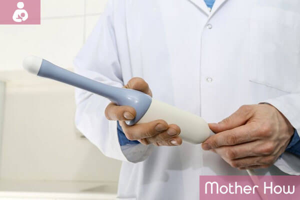
This gynecological examination of the uterus and other organs of the pelvis greatly simplifies the diagnosis, it can be done often, if necessary repeatedly. The sensor looks like a plastic rod about 12 cm long, its diameter is 3 cm, the handle is usually beveled, and at the end, there is a special channel with a needle for biopsy.
Indications for Transvaginal Ultrasound
Indication for this method of examination is a suspicion of pelvic organs diseases, urgent conditions (e.g., ectopic pregnancy), control of ongoing treatment, etc. Transvaginal ultrasound is performed under the following conditions:
- Diagnosis of pregnancy in the early stages
- Pain in the lower abdomen
- Diagnosis of the causes of infertility and monitoring the follicular apparatus of the ovaries;
- Violations of the menstrual cycle (delay in menstruation, bleeding in the middle of the cycle), pathological discharge from the genital tract
- Identification of inflammatory gynecological diseases
- Diagnosis of small pelvis neoplasms, including uterine myomas, endometriosis, ovarian cysts, etc.
- Taking hormonal drugs, the presence of intrauterine contraceptives (IUD) to monitor the condition of the endometrium and prevent complications
- Early pregnancy, when traditional transabdominal access (through the abdominal wall) is poorly informative
- Monitoring the procedure of IVF (in vitro fertilization)
- Determining the causes of urological diseases, urination disorders, urinary incontinence and urethral pathology
Transvaginal ultrasound of the pelvic organs is an ideal option for women with obesity since the usual study through the abdominal wall is not informative enough.
Precautions for the Diagnostics
When transvaginal ultrasound is performed, the following factors should be taken into account:
- The study is not carried out in the case when the patient has an allergic reaction to latex
- During the period of such manipulation, a patient should not move during the time indicated by the doctor
- For performing a vaginal ultrasound, the bladder should not be filled, except for the case when the patient is pregnant
Contraindications
There are no absolute contraindications to transvaginal ultrasound. The virgins can be examined through the rectum (transrectally). Transvaginal ultrasound during pregnancy is justified only in the early stages (up to 11-12 weeks).
Preparation for Transvaginal Ultrasound
For transvaginal ultrasound of the uterus and appendages, special preparation is not required. When you visit the ultrasound room, you will need a towel on which you will lie during the research.
If a transvaginal ultrasound is performed during pregnancy, the patient’s bladder should be moderately filled (drink about 500 ml of fluid one hour before the examination).
An obligatory condition for a transvaginal ultrasound of the pelvic organs is the absence of gas in the intestine. To do this, 2-3 days prior to the study, it is necessary to limit the products that cause increased gas production (vegetables, fruits, bread, dairy products, confectionery), and some drugs that reduce gas formation in the intestine (the enzyme, activated charcoal) are recommended.
There is no need to perform cleansing enemas before the study. Also, transvaginal ultrasound of the uterus and appendages is not necessarily performed on an empty stomach.
In emergency cases, transvaginal ultrasounds can be performed without preparation. But in this case, its informative value can be reduced.
Ultrasound examination of gynecological organs is recommended in the first half of the menstrual cycle (usually on the 5th-7th day), since in the second half the endometrium of the uterus is in the secretory phase, which can lead to incorrect interpretation of the results.
However, with endometriosis, transvaginal ultrasound of the uterus is recommended in the second phase of the cycle. To evaluate folliculogenesis (formation and development of ovarian follicles), the study should be carried out on the 5.9, 11-14, and 15 days of the menstrual cycle.
How is Transvaginal Ultrasound Performed?
You take off all the clothes below the waist, lay down on the couch, and bend your knees. In the same pose, any gynecological examination is performed.
The doctor puts a condom on the sensor and lubricates it with a special gel that performs two functions: eliminates the air space between the sensor and the organs, and serves as a lubricant for better penetration.
The vaginal sensor, or as it is also called a transducer, is gently and slowly injected into the vagina.
Due to the absence of sharp movements and a small depth of penetration, the procedure should not cause unpleasant and painful sensations in a woman.
The screen displays the organs that are being examined, the doctor records the necessary data. The procedure lasts no more than 5 minutes.
What Diseases Can This Procedure Diagnose?
This method of ultrasound scanning will help the doctor to determine exactly how well the female reproductive system, uterus, ovaries, and fallopian tubes are functioning. It is also often used to diagnose pregnancy pathologies. With such a scan, you can detect the following diseases and developmental features:
- Ovarian cyst
- Endometriosis
- Uterine and ectopic pregnancy
- Inflammatory processes
- The presence of pathological fluids – blood or pus in the fallopian tubes
- Uterine fibroids
- Polyposis of the endometrium
- Partial or full bladder skidding
- Different types of tumors: benign and malignant
- Chorionepithelioma
- Rupture of ovarian cysts or ovarian cancer
- Fluid in the small pelvis of a woman
Ultrasound, performed this way, also helps a woman to know when she is ready to conceive. For this, it is enough to follow the development of the ovaries. During the procedure, the doctor may inject a special contrast substance into the fallopian tubes, which helps to see whether they are passable. This method is indispensable in the treatment of infertility. Also, only transvaginal ultrasound is able to catch the child’s heartbeat at the time of 5 weeks of pregnancy.
Transvaginal Scanning During Pregnancy
This method is most often used on the first ultrasound in pregnancy, as the sensor is able to show the presence of the fetal egg within a few days after the delay in menstruation. Also, the transvaginal method of the ultrasound examination is the most informative with suspicion of the pathology of the development of the first stage of pregnancy. It will help to establish whether there is a threat of miscarriage, placental abruption and whether the thickness of the chorion is sufficient – a special inner layer of the uterus, from which the placenta is formed.

Preparation for such a study during pregnancy is no different from the preparation for an ultrasound of a non-pregnant woman, but keep in mind that this method is used only in the first trimester. At a later date, a conventional method of scanning through the abdominal cavity is used, since the transvaginal ultrasound can provoke contractions or cause the appearance of the uterine tone.
Complications of Transvaginal Ultrasound
If the procedure of transvaginal ultrasound is properly performed, there are no complications.
This examination is recommended to be performed even in healthy women for preventive purposes, at the age of up to 40 years it must be done at least once every 2 years, and after 40 years annually.

Born in Belarus, 1985, a pedagogue and family psychologist, mother. Taking part in procedures of social adaptation of the foster children in new families. Since 2015 is a chief editor of the motherhow.com project, selecting the best and up-to-date material for those, who are planning, expecting, and already having babies.